Slicer uniquely integrates several facets of image-guided medicine into a single environment. It provides capabilities for automatic registration (aligning data sets), semi-automatic segmentation (extracting structures such as vessels and tumors from the data), generation of 3D surface models (for viewing the segmented structures), 3D visualization, and quantitative analysis (measuring distances, angles, surface areas, and volumes) of various medical scans.
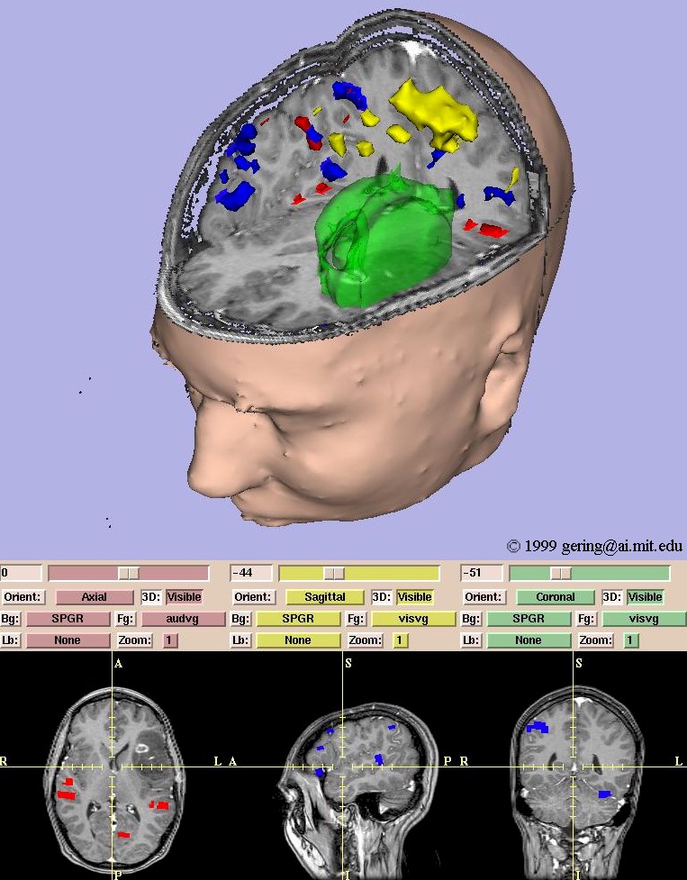 |
The image to the left was created by segmenting an anatomical MR scan to form 3D surface models of the skin and tumor (green). Functional MR data was segmented to build models of the motor cortex (yellow), auditory verb generation (red), and visual verb generation (blue). All these surface models are integrated in a 3D view along with 3 slices through the MR images. The slices are also shown in 2D views at the bottom. Note that the functional data is overlaid in color on the anatomical data. |
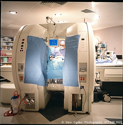
We integrated Slicer with an open MR scanner to augment intra-operative imaging with a full array of pre-operative data. The same analysis previously reserved for pre-operative data can now be applied to exploring the anatomical changes as the surgery progresses. Surgical instruments are tracked and used to drive the location of reformatted slices. Real-time scans are visualized as slices in the same 3D view along with the pre-operative slices and surface models. The system has been applied in over 20 neurosurgical cases at Brigham and Women's Hospital, and continues to be routinely used for 1-2 cases per week.
 |
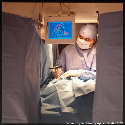 |
| The surgeon stands within the gap of the interventional MR magnet. (Signa SP -- GE Medical Systems) | The surgeon observes the display output of Slicer on the overhead LCD monitor. |