This is the archival of the developer site of the OpenKnee project - Generation 1. The development efforts for this generation of Open Knee were organized by Ahmet Erdemir and CoBi Core of the Cleveland Clinic. This study branched from a previous NIH funded study on multiscale modeling and simulation of the knee joint, J2C. For current activities (Generation 2), check FrontPage.
Previously:
-- aerdemir 2010-08-10 14:16:45 We are working on a User's and Developer's guide to prepare a tidy release of OpenKnee. Work on progress on this document can be found at https://simtk.org/websvn/wsvn/openknee/_gen1/doc/guide.odt in OpenOffice.org format.
-- -- siboles 2010-04-21 22:16:44 Python script, abq2feb.py and abq2feb.cnfg, is now available and can be downloaded at https://simtk.org/websvn/wsvn/openknee/_gen1/src
-- siboles 2010-10-05 22:19:34 New geometries have been generated from a reconstruction using ITK snap. These have been committed to the repository in a new directory ../dat/geo/version2geo/. Additions include: patella, patella cartilage, patella tendon, fibula, complete lcl, proximal and distal tibial mcl attachment, and meniscal horns. We are currently in the process of meshing these in TrueGrid this time with much more focus on the ability to edit mesh properties more easily.
Contents
Goals
- to provide a knee joint model (and related data) at various stages of its development
- for early testing and evaluation by any interested investigators
- to encourage collaborative work
- for reusability by others through open access
- as a learning tool for finite element analysis and knee biomechanics
Specific to our research interests:
- to develop a knee model representative of experimental overall joint response with the capability to predict cartilage stresses.
Roadmap
Releases relies on the following numbering scheme:
version.major.minor.revision
- version
- numbering based on goals of the roadmap
- major
- implementation of a new feaure
- minor
- improvement of a feature or a bug fix
- revision
- revision number of the subversion repository on which the release is based on
Version 1.0
- Imaging data; DICOM files
- Geometry; STEP or IGES files
- Mesh; text based input deck
- Literature based material properties
- Frictionless contact between tissue structures
- Loading and boundary conditions representative of passive flexion
- Output at desired time increments
Version 2.0
Long Term
Release Notes
Version 1.0
Team Roles
- Bhushan Borotikar (data collection; MRI and mechanical testing)
- Ton van den Bogert (supervision of data collection; knee biomechanics)
- Steve Maas (development and support for finite element analysis software; FEBio)
- Jeff Weiss (supervision of FEBio development; tissue mechanics)
- Craig Bennetts (initial geometry generation)
- Ahmet Erdemir (project oversight)
- Scott Sibole (technical lead and work on modeling and simulation procedures)
Specifications
Geometry
Source: https://simtk.org/websvn/wsvn/openknee/_gen1/dat/geo/
Currently, the knee geometry relies on manual digitization generated from sagittal MR images. Volsuite was used for this purpose. This initial geometry set was generated by Craig Bennetts of CoBi Core at Cleveland Clinic. 3D spline curves were used to develop NURBS surfaces using the loft feature in the CAD package Rhinoceros. Due to poor visibility of the lateral collateral ligament in sagittal image sets, its geometry is an approximation. Geometries are provided in a coordinate system aligned with the first sagittal MR image:
- origin - top-left corner
- x-axis - pointing towards right (anterior to posterior)
- y-axis - pointing downwards (superior to inferior)
- z-axis - pointing inwards (medial to lateral)
Mesh
Source: https://simtk.org/websvn/wsvn/openknee/_gen1/dat/msh/
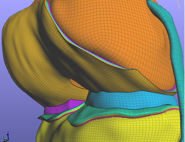
Current discretization relies on a fully hexahedral mesh generated using TrueGrid software (XYZ Scientific). The mesh file is a text based file conforming mesh definition convention of Abaqus.
A translator has been written to generate the complete model in FEBio with all materials, boundary conditions, and loads assigned. This also transforms the mesh into the widely used Grood & Suntay coordinate system. Grood & Suntay (1983)
- origin - most distal point on posterior femur midway between lateral and medial condyles
- x-axis - medial-lateral flexion axis
- y-axis - posterior-anterior of joint capsule
- z-axis - positive from distal femur to femoral head
Source: https://simtk.org/websvn/wsvn/openknee/_gen1/src
- python script: abq2feb.py
- configuration file: abq2feb.cnfg (comment with ##)

Element Sets:
- femur
- tibia
- femoral cartilage - fcart
- femoral cartilage w/o patella region - fcartr
- deep zone - fcartb
- transitional zone - fcartm
- superficial zone - fcartt
- tibial cartilage - tcart
- deep zone - tcartb
- transitional zone - tcartm
- superficial zone - tcartt
- medial meniscus - med meni
- lateral meniscus - lat meni
- medial collateral ligament - mcl
- anterior mcl - amc
- middle mcl - mmc
- posterior mcl - pmc
- lateral collateral ligament - lcl
- anterior lcl - alc
- middle lcl - mlc
- posterior lcl - plc
- anterior cruciate ligament - acl
- anterior acl - aac
- posterior acl - pac
- femoral insertion region (fibers defined by different nodal numbering due to need for different meshing technique) - aclfiber
- posterior cruciate ligament - pcl
- anterior pcl - apc
- posterior pcl - ppc
Surface Sets:
- femoral cartilage surface - fcs
- femoral cartilage surface w/o patella region - fcsr
- tibial cartilage surface - tcs
- femur surface contacting mcl - femmcl
- femur surface contacting lcl - femlcl
- tibia surface contacting mcl - mcltib
- tibia surface contacting lcl - lcltib
- mcl surface - mcl_surf
- only lateral side of mcl - mcls
- lcl surface - lcl_surf
- only medial side of lcl - lcls
- acl surface - aclsurf
- pcl surface - pclsurf
- lateral meniscus surface contact tibial cartilage - lmtib
- lateral meniscus surface contact femoral cartilage - lmfem
- medial meniscus surface contact tibial cartilage - mmtib
- medial meniscus surface contact femoral cartilage - mmfem
Node Sets
- Rigid interface nodes for femoral cartilage to femur attachment - f2fem
- Rigid interface nodes for tibial cartilage to tibia attachment - tc2tib
- Rigid interface nodes for ligament to femur attachment - femlig
- Rigid interface nodes for ligament to tibia attachment - tiblig
- Node set for anterior-lateral meniscal horn - lmant
- Node set for posterior-lateral meniscal horn - lmpost
- Node set for anterior-medial meniscal horn - mmant
- Node set for posterior-medial meniscal horn - mmpost
Anterior, middle, and posterior ligament regions were defined for pre-strain definition based on data from literature. Pena et. al. 2006
Material Properties
All material formulations in FEBio use a decoupled formulation splitting deviatoric and dilatational stresses/strains. We therefore dropped the conventional ~ over all decoupled quantities for convenience. The reader should just assume all quantities are in their decoupled form.
Bone
Rigid
Cartilage
Incompressible, isotropic Mooney-Rivlin: latex(\begin{displaymath}C_{1}=0.856\end{displaymath}), latex(\begin{displaymath}C_{2}=0.0\end{displaymath}), and latex(\begin{displaymath}K=20.833\end{displaymath}) (E=5 MPa, latex(\begin{displaymath}\nu=0.46\end{displaymath})) Li (2001)
Strain Energy Function: latex(\begin{displaymath}W=C_{1}(I_{1}-3)+C_{2}(I_{2}-3)+\frac{K}{2}(ln(J))^2\end{displaymath})
Young's modulus and poisson ratio from linear elastic model were converted to shear and bulk modulus using relationships: latex(\begin{displaymath}\mu=\frac{E}{2(1+\nu)}\end{displaymath}) and latex(\begin{displaymath}K=\frac{2\mu(1+\nu)}{3(1-2\nu)}\end{displaymath}) with latex(\begin{displaymath}C_{1}=\frac{\mu}{2}\end{displaymath})
**Note: By setting C2 to zero Mooney-Rivlin reduces to the neo-Hookean model. Mooney-Rivlin was used since FEBio does not have a decoupled implementation of neo-Hookean.
Poroelastic representation of cartilage mechanical response is a future extension possibility.
Ligament
Incompressible, transversely isotropic Neo-Hookean:
Strain Energy Function: latex(\begin{displaymath}W=C_{1}(I_{1}-3)+F_{2}(I_{4})+\frac{1}{2}K(ln(J))^2\end{displaymath})
where latex(\begin{displaymath}I_{4}\frac{\partial F_{2}}{\partial I_{4}} = 0 \ \ I_{4} \le1 \end{displaymath}) ; latex(\begin{displaymath}I_{4}\frac{\partial F_{2}}{\partial I_{4}} = C_{3}(e^{C_{4}(I_{4}-1)}-1) \ \ 1 < I_{4} <\lambda\end{displaymath}) ; latex(\begin{displaymath}I_{4}\frac{\partial F_{2}}{\partial I_{4}} = C_{5}+C_{6}I_{4} \ \ I_{4} \ge \lambda \end{displaymath})
latex(\begin{displaymath}I_{4} = \mathbf{a_{0}}\cdot\mathbf{C}\cdot\mathbf{a_{0}}\end{displaymath}) where latex(\begin{displaymath}\mathbf{a_{0}}\end{displaymath}) is the initial fiber direction.
latex(\begin{displaymath}C_{6}\end{displaymath}) is set to ensure C1 continuity in F2.
MCL and LCL: C1=1.44 MPa, C3=0.57 MPa, C4=48, C5=467.1 MPa, lambda=1.062, K=397 MPa Gardiner (2003)
PCL: C1=3.25 MPa, C3=0.1196 MPa, C4=87.178, C5=431.063 MPa, lambda=1.035, K=122 MPa Pena (2006)
ACL: C1=1.95 MPa, C3=0.0139 MPa, C4=116.22, C5=535.039 MPa, lambda=1.046, K=73.2 MPa Pena (2006)
An ongoing problem in modeling of the knee is the identification of ligament slack lengths (if ligaments were modeled as line elements) or zero stress-strain state of the ligament (which dictates in situ strain at reference model configuration). Current ligament modeling is aimed towards providing an adequate overall joint stiffness characteristics. Therefore, in future, it may be possible to use line elements to simplify their representation.
Meniscus
Fung Orthotropic Hyperelastic:
E1 = 125 MPa (circumferential)
E2 = 27.5 MPa (radial)
E3 = 27.5 MPa (inferior-superior)
latex(\begin{displaymath}\nu_{12} = 0.1 \end{displaymath})
latex(\begin{displaymath}\nu_{23} = 0.33 \end{displaymath})
latex(\begin{displaymath}\nu_{31} = 0.1 \end{displaymath})
G12 = 2 MPa
G23 = 12.5 MPa
G31 = 2 MPa
c = 1
K = 10 MPa
Strain Energy Function: latex(\begin{displaymath}W=\frac{1}{2}c(e^Q-1)\end{displaymath})
where,
E is the Green-Lagrange strain tensor
latex(\begin{displaymath}\mathbf{A_{a}}^0 = \mathbf{a_{a}}^0\otimes\mathbf{a_{a}}^0\end{displaymath}) is the material axis defined by the fiber's initial direction, latex(\begin{displaymath}\mathbf{a_{a}}^0\end{displaymath})
The orthotropic Lame parameters relate to Young's moduli, Poisson ratios, and shear moduli as follows:
latex(\begin{displaymath} \mu_{1}=G_{12}+G_{31}-G_{23} \end{displaymath})
latex(\begin{displaymath} \mu_{2}=G_{12}-G_{31}+G_{23} \end{displaymath})
latex(\begin{displaymath} \mu_{3}=-G_{12}+G_{31}+G_{23} \end{displaymath})
Interactions
Ligaments are attached to bone via rigid interface definitions (interface nodes become part of rigid body).
Frictionless, sliding contact defined between tibial to femoral cartilage, cartilage to meniscus, ligament to bone, and ACL to PCL.
Loading & Boundary Conditions
The loading should allow prescription of tibiofemoral joint flexion and application of loads to the remainder of 5 degrees of freedom of the joint.
Output
- Stress distribution
- Strain distribution
- Tibiofemoral joint kinematics (pose and orientation)
- Tibiofemoral joint kinetics (forces and moments)
- Contact stress
- Ligament forces
Solver
Non-linear system is solved using a standard BFGS quasi-Newton algorithm or full Newton method implemented by Steve Maas in FEBio. The linear system at each iteration is solved using Pardiso, a sparse matrix solver for shared memory architecture. http://www.pardiso-project.org/
Software
For finite element analysis FEBio, a freely accessible package, will be used. This software is a product of significant efforts by Jeff Weiss and his group from the Musculoskeletal Research Laboratories at the University of Utah. Current version used in this project is FEBio 1.2, which can be downloaded from their site.
Settings
Data
Data for model development efforts are courtesy of van den Bogert Laboratory at the Cleveland Clinic. The information was collected is part of doctoral work conducted by Bhushan Borotikar.
Specimen
NDRI ID |
08956 (Specimen acquired from National Disease Research Exchange) |
MRMTC# |
022508-03 (Specimen tested in Musculoskeletal Robotics and Mechanical Testing Facility at the Cleveland Clinic) |
Side |
Right |
Donor Age |
70 years |
Donor Estimated Body Weight |
170 lbs (77.1 kg) |
Donor Heigt |
5'6" (1.68 m) |
Donor Gender |
Female |
Donor Cause of Death |
Pneumonia/Cancer |
Imaging
Source: https://simtk.org/websvn/wsvn/openknee/_gen1/dat/mri/
The knee specimen was imaged at the Biomechanics laboratory of the Cleveland Clinic using a 1.0T (Tesla) extremity MRI scanner (Orthone, ONI Medical Systems Inc, Wilmington MA). The scanner has the capability to scan upper and lower extremities of up to 180mm diameter. A scanning protocol that gave a good contrast for articular cartilage and ligaments in the same scan were used Borotikar (2009). The specifics of this protocol are detailed in following:
Setting for Magneric Resonance Imaging |
|||
Scan Parameters |
|||
|
Sagittal |
Axial |
Coronal |
Pulse sequence |
GE3D |
GE3D |
GE3D |
TR |
30 |
30 |
30 |
TE |
8.9 |
8.9 |
8.9 |
Frequency |
260 |
260 |
260 |
Phase |
192 |
192 |
192 |
FOV |
150 |
150 |
150 |
BW |
20 |
20 |
20 |
Echo train |
1 |
1 |
1 |
NEX |
1 |
1 |
1 |
Flip angle |
35 |
35 |
35 |
Time |
5.03 |
3.19 |
3.30 |
Scan Options |
|||
|
Sagittal |
Axial |
Coronal |
Graphics SL |
Y |
Y |
Y |
RF spoiling |
Y |
Y |
Y |
Fat suppression |
N |
N |
N |
Minimum TE |
Y |
Y |
Y |
Inversion recovery |
N |
N |
N |
Partial data |
N |
N |
N |
No phase wrap |
Y |
Y |
Y |
Spatial saturation |
N |
N |
N |
Flow comp |
N |
N |
N |
Magnetic transfer |
N |
N |
N |
Prescan Parameters |
|||
|
Sagittal |
Axial |
Coronal |
Prescan |
Auto |
Auto |
Auto |
Center freq. |
Peak |
Peak |
Peak |
Slice Parameters |
|||
|
Sagittal |
Axial |
Coronal |
Number of slices |
70 |
45 |
60 |
Slice thickness (mm) |
1.5 |
1.5 |
1.5 |
Gap (mm) |
0 |
0 |
0 |
Range (mm) |
105 |
67.5 |
90 |
The knee was kept in full extension position. Imaging technique utilizes 3D spoiled gradient echo sequence with fat suppression, TR = 30, TE = 6.7, Flip Angle = 200, Field of View (FOV) = 150mm X 150mm, Slice Thickness = 1.5mm. Scans in three anatomical planes, axial, sagittal, and coronal, were conducted. Total scanning time was approximately 18 minutes. Selecting these specific sequence parameters produced images that highlighted articular cartilage such that it could be easily discriminated from surrounding bone and tissue. The protocols and the image set reflect partial data from the doctoral work of Borotikar (2009).
Mechanical Testing
Documentation
Source: https://simtk.org/websvn/wsvn/openknee/_gen1/doc/guide.odt
The source location includes a draft of the User's and Developer's guide in OpenOffice.org format.
Simulations
tf_joint.feb@88 - 100 N compressive load applied from t=0..1. 90 degree (1.57 radian) rotation then applied from t=1..4 with 100 N compressive load held constant. Job randomly terminated without an exit flag.
meniscectomy_45deg.cnfg@147 - 100N compressive load from t=0-1. 45 degree flexion t=1-2.5. Equilibrate from t=2.5-3.5. Meniscus-cartilage contact disabled.
tf_joint_45deg.cnfg@146 - 100N compressive load from t=0-1. 45 degree flexion t=1-2.5. Equilibrate from t=2.5-3.5.
Test Suite
- Troubleshooting
- Mesh convergence
Physiological
- Passive knee flexion
- Anterior/posterior laxity
- Internal/external rotation laxity
- Varus/valgus laxity
- Gait like
Support
For questions or reporting bugs/problems with Open Knee please use the forums found here.
References
Grood ES, Suntay WJ. A joint coordinate system for the clinical description of three-dimensional motions: application to the knee. J Biomech Eng. 1983 May;105(2):136-144.
Borotikar, Bhushan, Subject specific computational models of the knee to predict anterior cruciate ligament injury, Doctoral Dissertation, Cleveland State University, December 2009.
Gardiner JC, Weiss JA. Subject-specific finite element analysis of the human medial collateral ligament during valgus knee loading. J. Orthop. Res. 2003 Nov;21(6):1098-1106.
Peña E, Calvo B, Martínez MA, Doblaré M. A three-dimensional finite element analysis of the combined behavior of ligaments and menisci in the healthy human knee joint. J Biomech. 2006;39(9):1686-1701.
Li G, Lopez O, Rubash H. Variability of a Three-Dimensional Finite Element Model Constructed Using Magnetic Resonance Images of a Knee for Joint Contact Stress Analysis. J. Biomech. Eng. 2001;123(4):341-346.
Jiang Yao et al., Stresses and strains in the medial meniscus of an ACL deficient knee under anterior loading: a finite element analysis with image-based experimental validation. J, of Biomech. Eng. 2006;128(1):135-141.
Literature on Finite Element Representation of the Knee Joint
Results of a search with keywords ("finite element" AND knee) can be accessed:
http://www.ncbi.nlm.nih.gov/pubmed?term=%22finite%20element%22+knee
Review of finite element representation of the knee joint can be found in /LiteratureReviewKneeFea