Contents
SMULTIS001-1 mock
- donor: CMULTIS002
- First mock of instrumented surgical tool procedures minus Vic3D strain measurement.
- Cut skin 10 cm - measured length, width, depth
- retracted skin 2 cm
- widened incision with forceps
- closed incision for approximation with forceps
- everted tissue with forceps
- (trial #16) closed suture with square knot at midpoint connected to scalpel bolt
- (17) closed suture with a loop, no knot, take first trial not last.
- (18) closed suture with surgeons knot (first knot)
- (19) tied 3rd knot (second was not instrumented)
(20 & 21) tissue pulling. Not a loop like (17). tied knot to one end, pulled on other. mechanically the same as the loop, just pulled on one side instead of both.
- Notes:
- finalize trial naming convention
- build in length, width, depth window after each trial
- build apparatus to hold metal ruler (use manfrotto arm)
SMULTIS001-2 mock
- donor: CMULTIS002 (the name was actually SMULTIS002-1 but this will be archived as mock data to leave 002 for a different donor)
Goal: Test Vic3D system integration and use during first three surgical procedures
- all software and hardware modifications were made prior
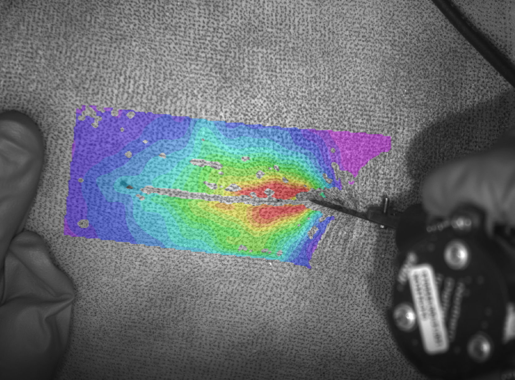
- Vic3D triggered at 30fps for 20s by multis computer
- Synchronization signal also sent and stored with Vic3D
- For each trial, a folder was created for Vic3D
- An ink roller was used and worked seemingly well
- Different camera angles were tested for optimal viewing and minimal tool shadowing
- Notes for next time
- Create calibration folder for the day's experiment
- use for all trials that day
- tilt cameras towards operator
- move cameras closer together to reduce shadowing effect
SMULTIS003-1 mock
- donor: CMULTIS003
Goal:
Full data collection including strain measurement
Notes:
- although DIC can only measure 4-5cm cut length, make cut full length. DIC will occur on first half, so start cut at beginning of DIC region, continue 5cm after end of DIC
- do not retract after skin cut with current retractor because it is too large relative to the cut depth (0.6mm)
- move the forceps wid/eve/clos and suture steps to after the cut through skin fat interface so that more tissue is available. The forceps will essentially be grabbing the skin plus skin/fat interface
- for retraction, start at midline and continue to retract 1cm after contact
- subtract ruler thickness from caliper depth measurement

SMULTIS004-1
- donor: CMULTIS004 date: 08-23-18
Goal:
First non-mock experiment
Protocol Deviations
- Specimen underwent 1 extra freeze thaw cycle due to software malfunction on first attempted experiment
- To reduce reflection during indentation the polarized VIC3d light filters were first optimized to the undeformed skin, then rotated 90 degrees. This method will be used from now on.
- A shadow not visible to the naked eye blocked VIC3d strain measurement of the cut trial. A mock cut will be performed from now on.
- Extra force/moment data was collected at the beginning of each trial. A series of tap tests were conducted to show that the extra data is exactly the length of the difference between the force data time (~13s) and optotrak data time (~10s). The last 10s of each optotrak and force are synchronized by removing that difference.
SMULTIS005-1
- donor: CMULTIS005 date: 09-14-18
Protocol Deviations
- missing opto data after incision completed in trial 24
SMULTIS006-1
- donor: CMULTIS006 date: 09-21-18
Protocol Deviations
- some missing optotrak data (beginning only) for trial 017 (cutting fat muscle interface)
SMULTIS007-1
- donor: CMULTIS007 date: 10-02-18
Protocol Deviations
- White ink used successfully for dark skin
SMULTIS008-1
- donor: CMULTIS008 date: 10-05-18
Protocol Deviations
- White ink used successfully for dark skin
SMULTIS009-1
- donor: CMULTIS009 date: 10-17-18
Protocol Deviations
- SXX_CUT_FAT
- Just over half the length of the cut penetrated the muscle fascia below
- SXX_CUT_FMI was therefore not possible and not collected following the SXX_CUT_FAT trial.
- The remaining fascia and roughly 3mm (depth) of muscle was cut but not recorded to prepare the tissue for the muscle retraction trial.
Notes
- Possibly best VIC3D yet for each of the three trials
SMULTIS010-1
- donor: CMULTIS010 date: 10-18-18
Notes
- VIC3D results for all three trials comparable to 009.
- Tissue recovered atypically well during indentation, likely due to excess and relatively young subcutaneous tissue
SMULTIS011-1
- donor: CMULTIS011 date: 10-30-18
Notes
- VIC3D cut terial was not the best. Tissue was not dark or light. Both white and black speckle was attempted
- This specimen underwent one less freeze-thaw cycle and was kept thawed one day less than others. The Ultrasound and Surgical Tools and CT/MRI were all completed in one day.
- Analysis:
- Trial 021 - missing Optotrak data in the middle of the dissection. Trial not repeated since dissection is destructive.
SMULTIS012-1
- donor: CMULTIS012-1 date: 11-06-18
- Very good VIC3D trials.
- Missing fat cut trial because of extremely thin fat layer.