|
Size: 6603
Comment:
|
Size: 6608
Comment:
|
| Deletions are marked like this. | Additions are marked like this. |
| Line 26: | Line 26: |
| = Final MRI Specifications = | = Final Imaging Specifications = |
Contents
Prerequisites Protocols
Initial Goal
A set of magnetic resonance (MR) images of the extremity specimen (upper/lower leg, upper/lower arm) with clear visual delineation of skin, fat, and muscle layers.
Additional information about the imaging facility, all associated contacts, and MRI hardware can be found at:
Position/Orient Specimen in MRI Machine
- Anatomical position, anterior up. Foot towards distal end of MRI.
Acquire Image Sequences
For each segment (upper leg, lower leg, upper arm, and lower arm), the following set of image collection protocols will be performed. All images should be acquired in the same coordinate system to be able to align reconstructed tissue geometries during assembly of the full segment geometry. To accomplish this the origin (isocenter) and the axes of the magnet, which is set at the beginning of the session, should not change. In addition, the specimen should not be moved. It should be noted that a pixel-by-pixel alignment of image sets (co-localization) is not necessary.
[Examples to come] Before, each sequence is acquired, verify that the acquisition properties match the desired settings in the tables below. After each sequence is acquired, inspect the images and compare them against the sample images below to to ensure that it was collected properly. If not, the sequence should be recollected.
Final Imaging Specifications
Sample Image Acquisition Properties:
|
T1 w/o Fat Sup |
T2 w/ Fat Sup |
Proton Density |
Plane |
Axial |
- |
- |
No. of slices |
25 |
- |
- |
No. of Seq/Segment |
4 |
- |
- |
FOV (mm) |
240 x 150 |
- |
- |
Slice thickness/gap (mm/mm) |
2/1 |
- |
- |
No. excitations averaged |
1 |
- |
- |
Distance factor (%) |
100% |
- |
- |
X-resolution (mm) |
0.47 |
- |
- |
Y-resolution (mm) |
0.47 |
- |
- |
Scan Time / Seq. (min.) |
3-4 |
- |
- |
Imaging Trial 4: 11/30/16
Attendees: Tyler, Rici, Ben (CC), Josh (UH)
SETTINGS 1: Upper Leg
This imaging protocol is similar to Settings 2 - Imaging Trial 2; to acquire axial plane image sets. The in-plane resolution was changed to approximately 0.43 mm and the field of view to 250mm . The out-of-plane resolution remained the same at approximately 3 mm (2 mm slice thickness and 1 mm gap).
The goal is to reduce the shadowing/darkness that appears at the beginning and/or end of a segment. Where a segment is a set number of slices. To achieve this, we tried several different protocols.
Sequence #1: 50 slices each segment - no overlap
Sequence #2: 50 slices each segment - overlap 10 slice
Sequence #3: 25 slices each segment - no overlap
The reasoning for sequence 3, reducing the slice number, is that the number of slices per segment can influence the image quality, in particular contrast.


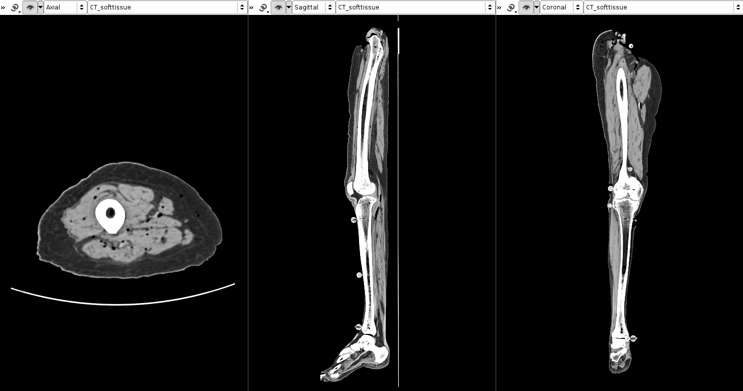
Imaging Trial 3 (CT): 11/29/16
Attendees: Tyler, Bong-Jae
SETTINGS 1: Upper Leg
The goal of this imaging session is to provide a set of Computer Tomography (CT) images to compare to MR images of the same specimen used in Imaging Trial 2. Specifically, we want to see if any of the tissue region boundries (skin, fat, muscle) can be more easily delineated.
Imaging Trial 2: 11/16/16
Attendees: Tyler, Ahmet, Rici (CC), Josh (UH)
SETTINGS 1: Upper Leg
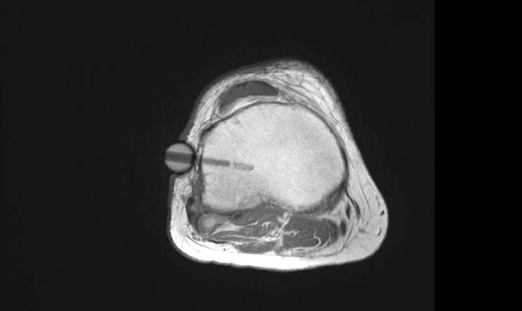
The goal of this imaging protocol is to acquire axial plane image sets with an in-plane resolution of approximately 0.47 mm, an out-of-plane resolution of approximately 5 mm (2.5 mm slice thickness and 2.5 mm gap) and a large enough field of view inclusive of the entire axial section of the specimen.
Sequence #1: PD type - axial plane - Proton Density
Sequence #2: T1 type - axial plane - WITHOUT Fat Suppression
Sequence #3: T1 type - axial plane - WITH Fat Suppression
SETTINGS 2: Lower Leg
The goal of this imaging protocol is to acquire axial plane image sets with an in-plane resolution of approximately 0.47 mm, an out-of-plane resolution of approximately 3 mm (2 mm slice thickness and 1 mm gap) and a large enough field of view inclusive of the entire axial section of the specimen.
Sequence #1: T1 type - axial plane - WITHOUT Fat Suppression
Imaging Specifications
Sample Image Acquisition Properties:
|
Settings 1 |
Settings 2 |
Imaging Date |
11/16/2014 |
11/16/2014 |
Plane |
Axial |
|
No. of slices |
50 |
50 |
No. of Seq/Segment |
3 |
4 |
FOV (mm) |
240 x 250 |
240 x 150 |
Slice thickness/gap (mm/mm) |
2.5/2.5 |
2/1 |
No. excitations averaged |
1 |
|
Distance factor (%) |
100% |
|
X-resolution (mm) |
0.47 |
|
Y-resolution (mm) |
0.47 |
|
Scan Time / Seq. (min.) |
3-4 |
|
Imaging Modality |
Proton Density |
T1 |
|
T1 |
|
|
T1 Fat Suppression |
Proton Density - Tibia |
|
T1 - Tibia |
|
T1 - Fat Suppression - Tibia |
|
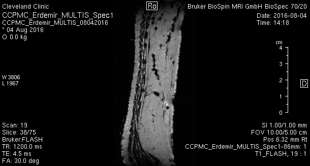
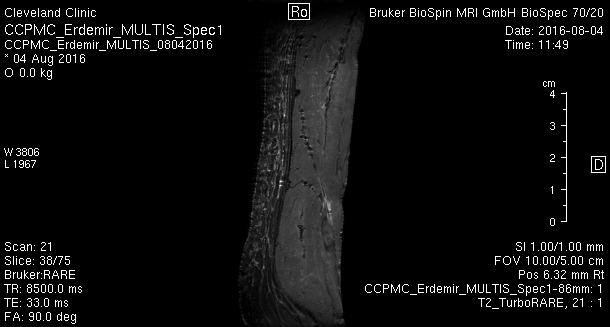
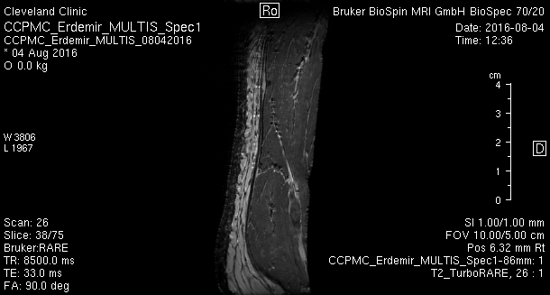
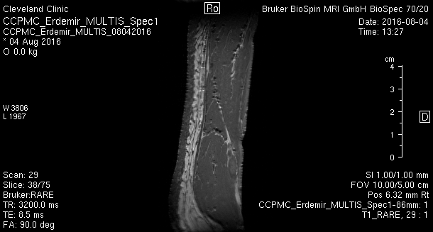
Imaging Trial 1: 8/4/16
- Imaging was performed in the LRI Animal MRI in ND1-09
- "Without fat suppression" is preferred, specifically scans
- 17, 18, 26, 29
. .
.
. .
.
. .
.
. .
.
.